Technology
To give you the healthy, beautiful, straight smile you've always wanted, Dr. Rosenthall utilizes innovative digital treatment planning. Using this digital technology, he can create an orthodontic treatment plan that is specifically customized for you.
Each patient's orthodontic condition is unique, which is why we develop each patient's treatment plan based on his/her specialized diagnosis. The treatment planning process involves taking digital photographs, X-rays and digital impressions.
Digital Photography
We utilize high-resolution digital photography to capture detailed images of your mouth, thus enabling us to maintain sharp, accurate records and keep you thoroughly informed of your treatment progress.
Digital X-rays
Digital X-rays, or radiographs, are also used in our office to provide us with invaluable information about your oral and dental health. By placing a wireless sensor in your mouth and using high-tech computer software, we are able to generate digital images of your mouth significantly faster than traditional X-rays. Not only are they friendly to the environment, they are much safer than traditional X-rays, reducing your exposure to radiation by 90 percent!
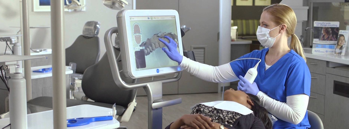
Digital Impressions/Models
We also utilize a digital impression system, which replaces the unpleasant-tasting, messy and sometimes inaccurate traditional putty impressions. Using a digital scanner, we will take three-dimensional (3-D) digital images, or impressions, of your teeth and bite. These impressions are then used at a laboratory where they will custom-fabricate a model of your teeth as well as customize bracket trays to increase the accuracy of your treatment.
All of these diagnostic records help us formulate a personalized treatment plan that meets the patient's precise needs and desires. By using this advanced technology, we are able to keep you up-to-date on your treatment progress by comparing your mouth to what it looked like before your treatment began.
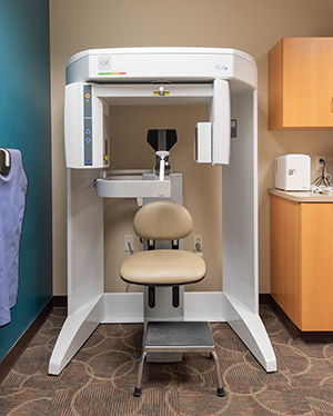
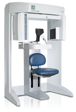 While CT (computed tomography) imaging has been used in the medical field for over 30 years, it is becoming the new diagnostic tool of choice for orthodontic analysis, diagnosis and treatment planning due to the latest advancements in diagnostic technology such as the revolutionary i-CAT® Cone Beam 3-D Imaging System. This three-dimensional CT technology can provide a quicker full scan of the head than traditional two-dimensional imaging, allowing orthodontists a better visualization of the hard and soft tissues of the craniofacial structures from several perspectives. In addition to the advantages of traditional CT scans, the i-CAT® Cone Beam releases up to 90% less radiation than traditional X-ray machines, enhancing your safety while providing crisp, clear images for more efficient diagnostic analysis and treatment. We feel our modern, cutting-edge techniques ensure you are receiving the quality care you deserve.
The i-CAT® Cone Beam 3-D Imaging System can immediately produce three-dimensional images in under one minute. This in-office, easy-to-use system provides your orthodontist a comprehensive view of all oral and maxillofacial structures, dramatically increasing the efficiency with which your orthodontist is able diagnose your condition and plan for your treatment.
i-CAT® technology is used for:
- TMJ assessment
- Surgical planning
- Assessment of cleft lip and palates
- Assessment of the alveolar bone
- Impacted tooth position
- Facial analysis
- Tongue size and posture
- Airway assessment
- Placement of dental implants